Visualizing Electric and Magnetic Field Coupling in Au-Nanorod Trimer Structures via Stimulated Electron Energy Gain and Cathodoluminescence Spectroscopy: Implications for Meta-Atom Imaging
Researchers from the University of Tennessee, Oak Ridge National Lab, the University of Washington, and Clarion University of Pennsylvania used the Waviks' Vesta to explore the near-field plasmonic responses of trimer meta-atoms composed of gold nano rods. Their work was published in ACS Applied Nano Materials. Their results open the possibility to explore the nanoscale excited-state near-field imaging of other magnetic meta-atom structures.

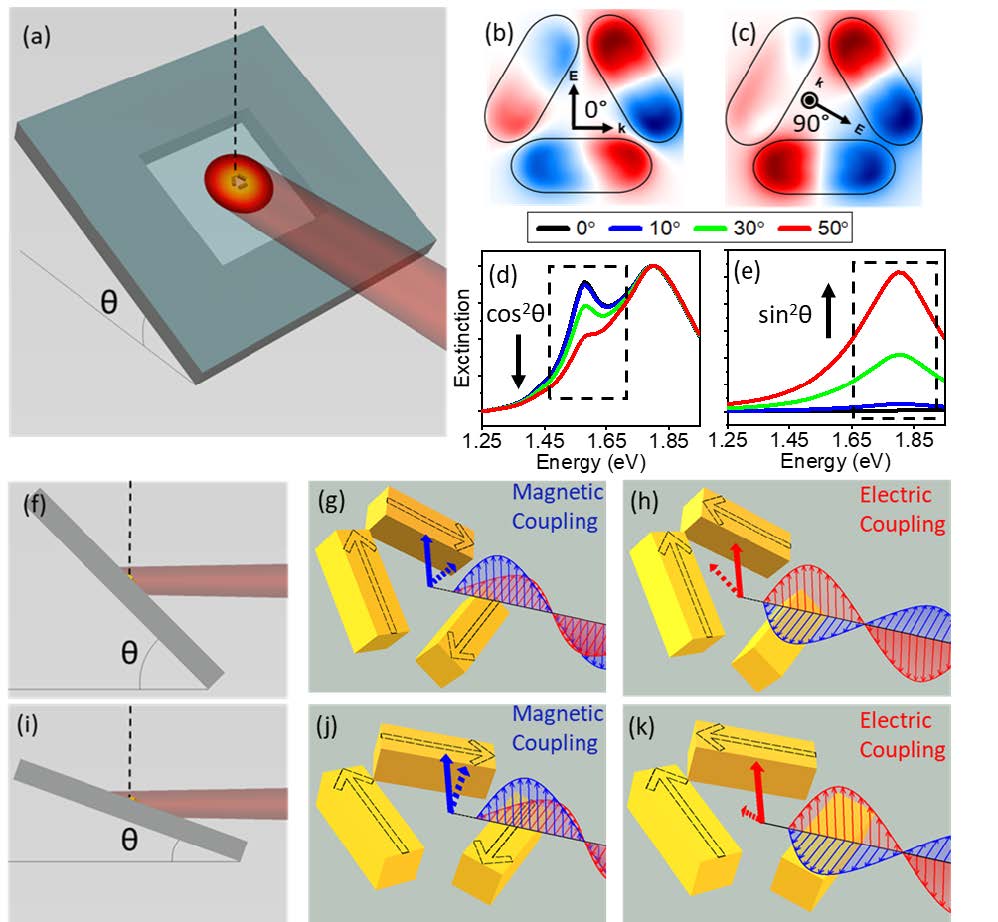
Figure 1: a) Schematic of the sEEGS experimental setup, θ indicates the sample tilt from normal to the electron beam (shown as a dotted line in a, f and i), b-c) calculated induced electric field maps of the magnetic and electric trimer modes with optical field orientations included as well as the laser-substrate angles relating the orientations to the plots directly below. In both cases, the normal component of the field at any integer multiple of the period is displayed (d) and electric mode under optical excitation (e), f-k) show the influence of sample tilt on the optical coupling of the magnetic (g and j) and electric (h and k) modes. Black arrows indicate the direction of the individual nanorod dipole oscillation relative to the other nanorods. Red arrows are used to show the electric field, while blue is used for the magnetic field of the photon. Dotted red and blue arrows indicate the projection of the electric and magnetic components onto the trimer substrate plane that it is operative.
The teams from the University of Tennessee, Oak Ridge National Lab, the University of Washington, and Clarion University of Pennsylvania demonstrated that stimulated electron energy gain spectroscopy is capable of probing and visualizing the near-field of electrically and magnetically driven plasmonic meta-atoms.
Reference:
Visualizing Electric and Magnetic Field Coupling in Au-Nanorod Trimer Structures via Stimulated Electron Energy Gain and Cathodoluminescence Spectroscopy: Implications for Meta-Atom Imaging
DOI: 10.1021/acsanm.1c03171
